
The use of computed tomographic (CT) techniques to create detailed, three-dimensional images of fossils has exploded in the past decade.
Using CT techniques, palaeontologists can look at the internal anatomy of fossil organisms in astounding detail without the risk of damaging them. CT data has provided unprecedented insights into the ecology and evolution of extinct species.
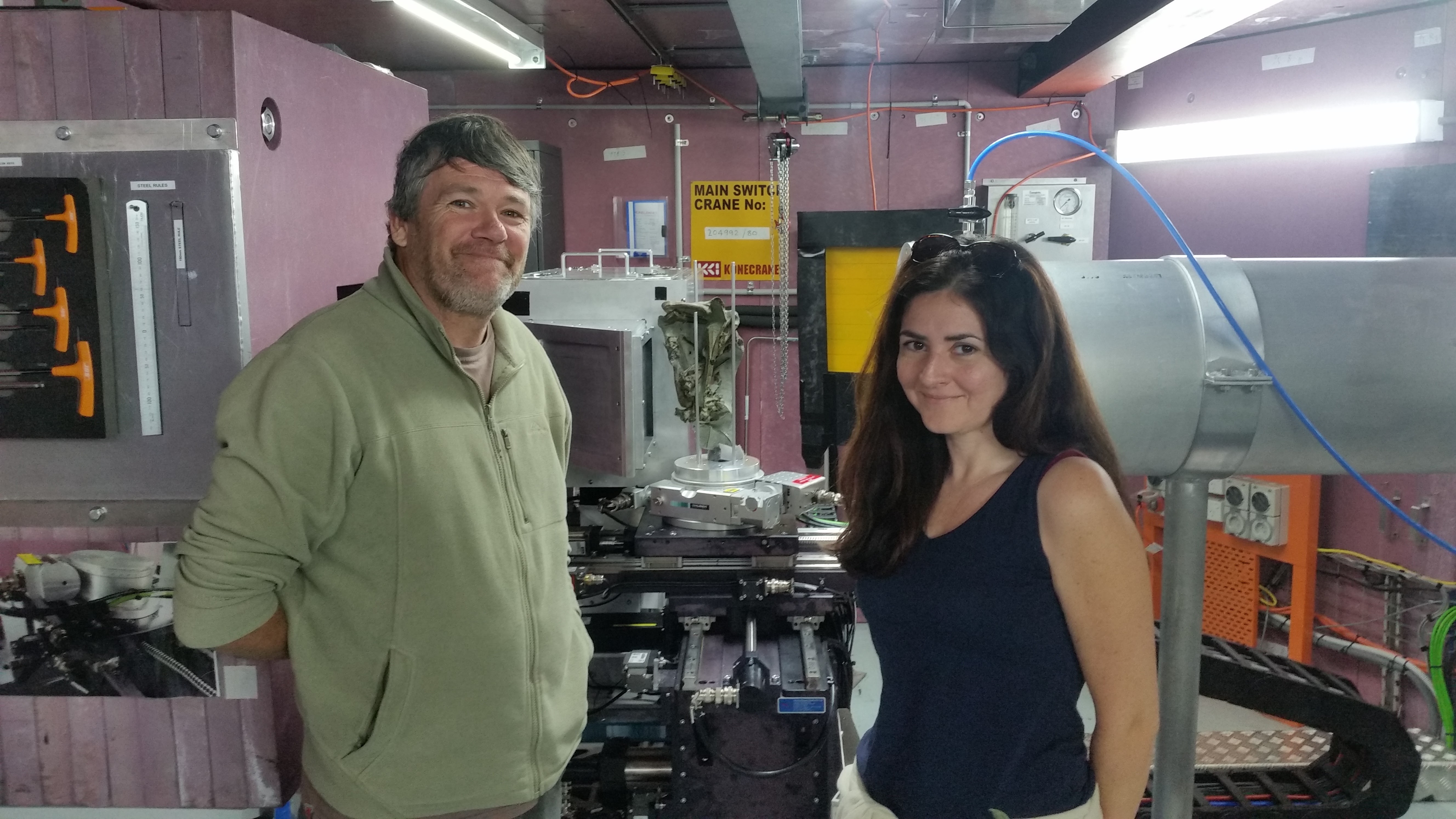
In November 2017 we were awarded a grant to carry out 3 days of research at the only operational nuclear reactor in Australasia. We wanted to use CT techniques to visualise and “virtually reconstruct” some of the world’s oldest fossil penguins.
The specimens, two from the Chatham Islands and one from Waipara, North Canterbury, were embedded in hard rock so it would be extremely difficult to get them out intact. These fossils are between 60 and 65 million years old, and are three of no more than 10 known fossil penguins this age worldwide. They could shine a light on the early stages of penguin evolution, not long after the disappearance of dinosaurs and other creatures.
The OPAL reactor at the Australian Nuclear Science and Technology Organisation (ANSTO) is in Lucas Heights, a suburb south of Sydney, New South Wales. OPAL is a research reactor. It generates 20 megawatts of heat using around 30 kg of uranium. This is very small compared to a typical nuclear power reactor, which may operate at 3,000 megawatts and contain around 100,000 kg of uranium.
For our work we used the neutrons that are fired out of the reactor as a by-product of nuclear fusion using an instrument called DINGO (named after the Australian mammal Canis lupus). DINGO works by "firing" neutrons at an object through a beam tube from the reactor. Some of the neutrons are absorbed by the object while others pass straight through. A camera on the opposite side of the object is able to capture images of the shadow projected as the object is slowly rotated around 360 degrees. These separate 2D images are then fitted together and turned into a 3D image. This process is called neutron tomography.
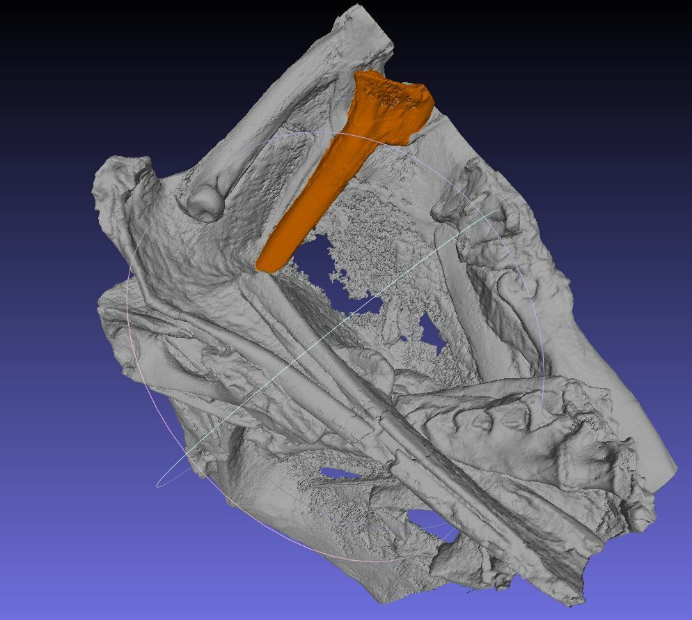
The images produced by neutron tomography look quite similar to the more common x-ray CT scans, but there are some important differences. X-rays work best when detecting dense material within lighter materials (like bone inside soft tissue or metallic weapons in hand luggage). Neutron scanning, on the other hand, is sensitive both to changes in density and to different minerals in rocks. The high energy of the neutrons enables them to “see” through very dense rock and especially through metals such as pyrite which are common in fossils from the North Canterbury area. This greater penetration is a major advantage when studying relatively large objects or specimens.
Neutron tomography produces a large amount of radiation, meaning the objects being scanned often have an afterglow and need to "cool" for hours or even days in a secure area until the radiation dissipates. The technique is non-destructive and the mineral samples can then be returned to museums unchanged, without the loss of any environmental information or soft tissue remains that may be contained within the rock surrounding the fossils.
Now that we have the images work is continuing to describe these remarkable early penguins. The experiment has also been useful in developing the techniques of neutron tomography. We have learnt that some rocks work well and some don’t and that these astounding new techniques will be useful in the future when collecting and preparing our unique fossil heritage.


