
A Health New Zealand Christchurch medical physicist has uncovered new evidence that could flip the origin story of New Zealand’s first X-ray on its head.
Until now, Dr William Hosking has been credited with taking the first X-ray in New Zealand in 1896, using what was then known as Röntgen rays. This device was named after Wilhelm Röntgen, a German physicist who first discovered the X-ray in 1895.
However, after months of careful sleuthing, Steven discovered that Dr Hosking’s ray machine didn’t arrive in the country until early 1897, which means the honour for the first X-ray goes to Augustus Hamilton, the registrar at the University of Otago.
Hamilton took his image, of a frog, before 12 May 1896, for a lecture on Röntgen rays by University of Otago colleague, Professor John Shand, Steven discovered. That was only six months after the initial discovery of X-rays, which is remarkably soon considering that shipping and communications took more time to reach New Zealand in the nineteenth century.

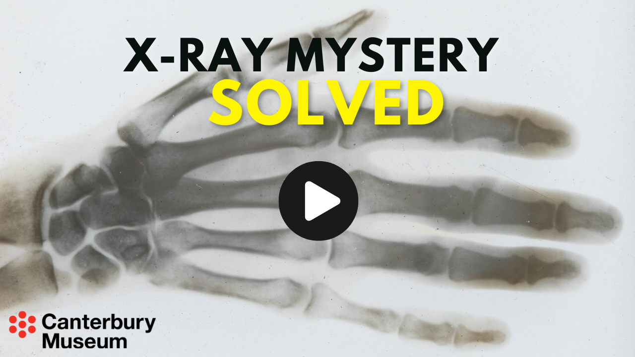
Sadly, Hamilton’s X-ray isn’t thought to have survived, but Steven's further research found New Zealand’s earliest known surviving X-ray, also of a frog, in Canterbury Museum.
Frogs were common subjects at the time, and this image was taken in September 1896 by Dr William Evans, who taught at Christ’s College before lecturing in chemistry and physics at Canterbury University College. Evans sourced the specimen from Museum Curator Frederick Hutton.
Steven worked alongside Canterbury Museum Associate Curator Human History, Frances Husband, to find the image in the Museum collection. Their detective work also found the original frog that the X-ray was taken from, which has been stored in preservative for 142 years. The frog – Leiopelma hochstetteri – came into the collection in 1883.
The Museum’s long-term project to inventory the entire collection of 2.3 million objects was key to making this discovery, particularly the Lotteries-funded inventory of 200,000 photographic negatives,
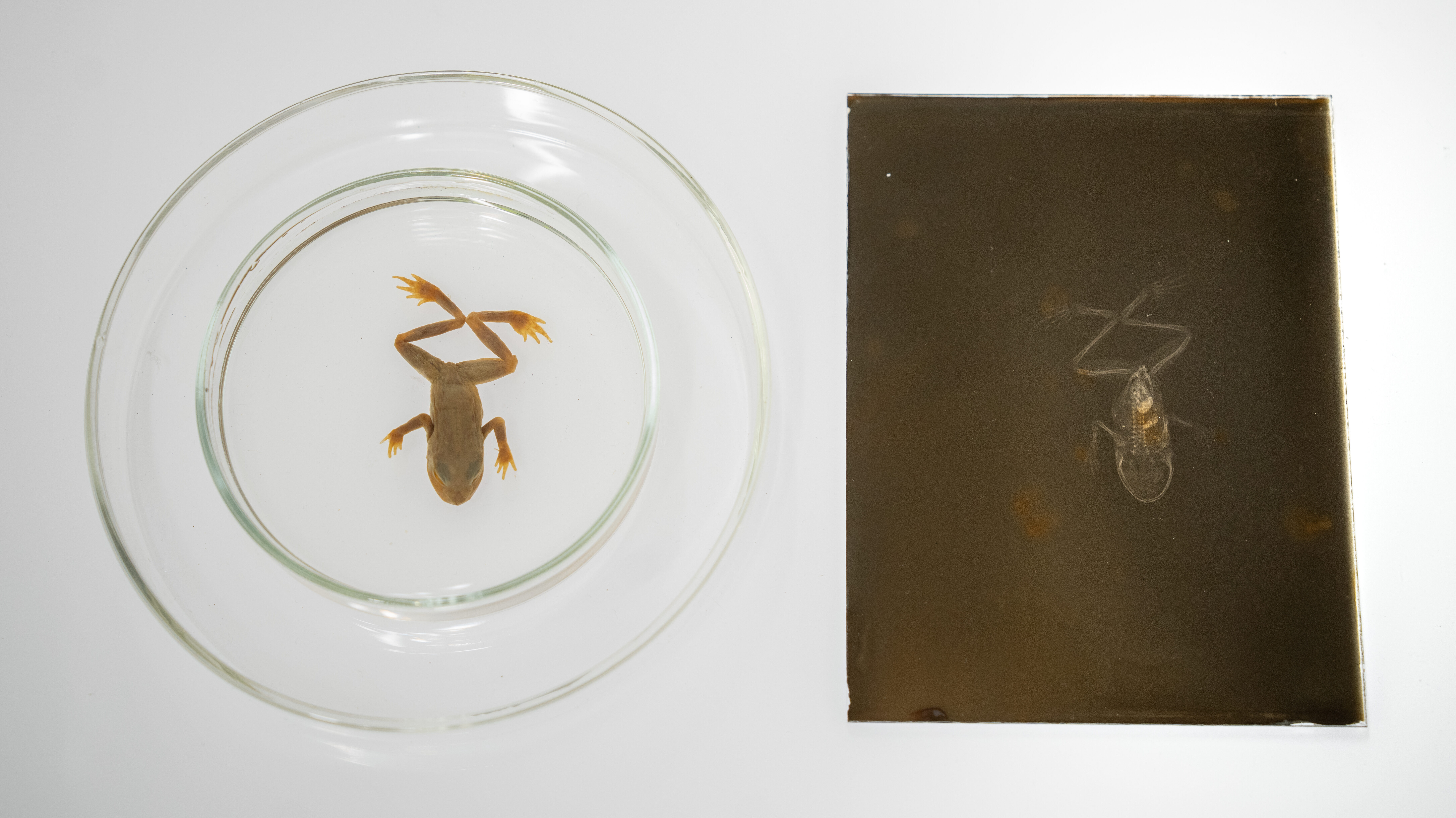
To make sure that the specimen found in the collection was the same frog, another X-ray was taken on the latest 2D and 3D high-resolution machines at Christchurch Hospital, after hours. The old and new X-rays were a perfect match.
Steven also worked with staff at Christ’s College school to locate the original X-ray tube that the 1896 image was taken with. The tube was found in a box in the back of the physics laboratory.
Steven presented these findings to his peers at the Grand Rounds health research lecture series today, just three days shy of the 130th anniversary of Rontgen’s discovery on 8 November.
Other significant findings from Steven’s research are planned for publication in conjunction with the Cotter Medical History Trust and include details on 15 early X-ray pioneers in New Zealand, nine of whom have never before been mentioned in histories of the invention in New Zealand.


